Diagnostic Mammogram
What Is The Difference Between A Diagnostic Mammogram And A Screening Mammogram?
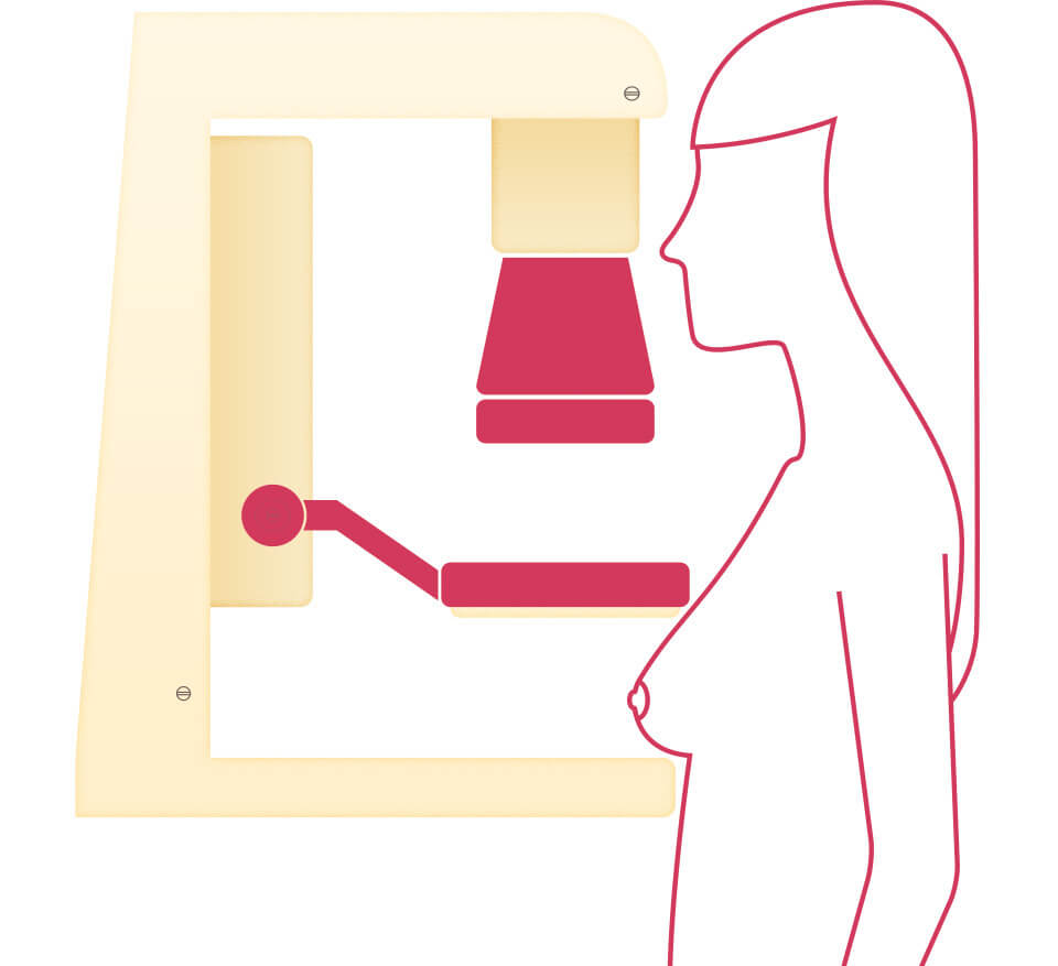
A mammogram is an x-ray of the breast. While screening mammograms are routinely performed to detect breast cancer in women who have no apparent symptoms, diagnostic mammograms are used after suspicious results on a screening mammogram or after some signs of breast cancer alert the physician to check the tissue.
Such signs and symptoms may include:
- A lump
- Breast pain
- Nipple discharge
- Thickening of skin on the breast
- Changes in the skin texture, such as dilation (or enlargement) of the pores of the breast
- Changes in the size or shape of the breast

A diagnostic mammogram can help determine if these symptoms are indicative of the presence of cancer. As compared to screening mammograms, diagnostic mammograms provide a more detailed x-ray of the breast using specialized techniques. They are also used in special circumstances, such as for patients with breast implants.
Beginning in late 2024, radiologists will be required to report the degree of density found within your breast tissue. Highly dense breast tissue is considered a risk factor for developing breast cancer. Having dense breast tissue can also make mammogram images harder to read and interpret. If you have dense breast tissue, you should discuss additional imaging studies, such as ultrasound or breast MRI, with your healthcare provider.
Do you have dense breasts?
Learn more about breast density and how it can affect your breast health in the free Dense Breasts Q&A Guide.
Dense Breasts Q&A GuideWhat’s Involved In A Diagnostic Mammogram?

If your doctor prescribes a diagnostic mammogram, realize that it will take longer than a normal screening mammogram, because more x-rays are taken, providing views of the breast from multiple vantage points. The radiologist performing the test may also zoom in on a specific area of the breast where there is a suspicion of an abnormality. This will give your doctor a better image of the tissue to arrive at an accurate diagnosis.
In addition to finding tumors that are too small to feel, mammograms may also spot ductal carcinoma in situ (DCIS). These are abnormal cells in the lining of a breast duct, which may become invasive cancer in some women.
These abnormal cells do not appear as a mass at all. Instead, they look like tiny grains of sand called microcalcifications. If these microcalcifications are grouped together and/or are in a row, this is a sign they might be DCIS. Not all DCIS findings progress into invasive cancer. There are studies currently being done to help determine which DCIS findings turn into invasive cancer to help physicians plan what treatment is best for a woman’s specific findings of DCIS inside the duct of the breast.
How Reliable Are Mammograms For Detecting Cancerous Tumors?
The ability of a mammogram to detect breast cancer may depend on the size of the tumor, the density of the breast tissue, and the skill of the radiologist performing and reading the mammogram. Mammography is less likely to reveal breast tumors in women younger than 50 years than in older women. This may be because younger women have denser breast tissue that appears white on a mammogram. Likewise, a tumor appears white on a mammogram, making it hard to detect.

There have been wonderful improvements in the last 20 years regarding mammogram technology. Today, it is best to get a 3D mammogram also known as tomosynthesis. This type of modern mammogram machine detects breast cancer 28% more accurately than older X-ray analog mammograms.
You can call our mammography facility beforehand to find out if they perform 3D mammography. You may also ask if the radiologist is a breast imaging radiologist. This also contributes to getting an accurate reading of your mammogram.
If you had prior mammograms done at a different facility, get those mammograms either sent to the new facility where you are going or pick them up yourself and take them there. It is important for the radiologist to always compare prior mammograms to the most recent one.
Materials on this page are courtesy of:


